Diagnosis and Management of Hepatic Hydrothorax

Learning Objectives
- Describe the pathophysiology and typical presentation of hepatic hydrothorax.
- Interpret pleural fluid studies and recognize spontaneous bacterial empyema.
- Summarize treatment options and current MELD exception guidance.
What is hepatic hydrothorax?
Hepatic hydrothorax (HH) is a pleural effusion related to portal hypertension in the absence of another primary cardiac, pulmonary, renal, pleural, or malignant cause. It occurs in approximately 4%–12% of patients with cirrhosis and is usually right-sided. Although most patients have ascites, it may not be clinically apparent. Patients can present with dyspnea, cough, pleuritic discomfort, or hypoxemia. HH is an important marker of decompensated cirrhosis.
Why does fluid collect in the chest?
Portal hypertension promotes sodium and water retention and ascites formation. Fluid moves through small defects in the diaphragm, drawn into the pleural cavity by negative intrathoracic pressure. These defects occur more often on the right, explaining the typical laterality. Ascites may be difficult to detect when fluid moves into the chest as quickly as it forms.
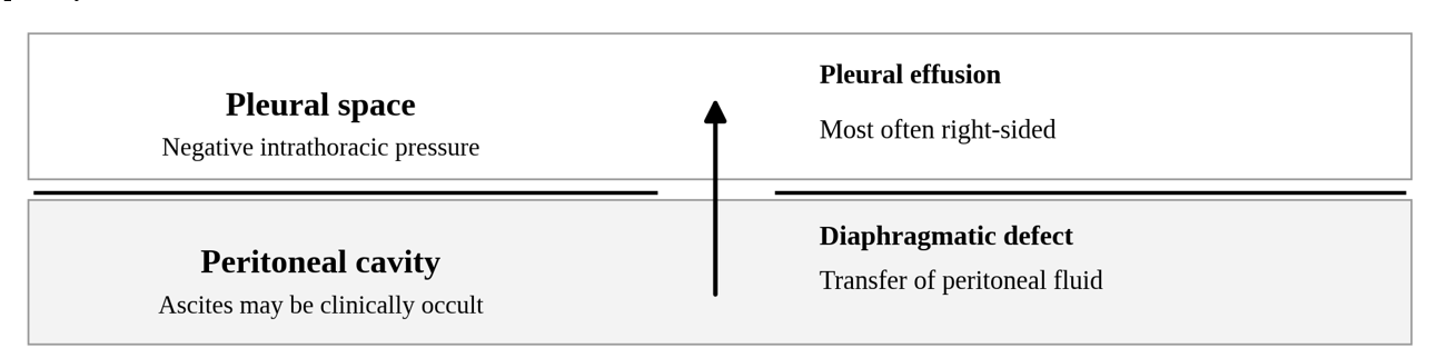
Figure 1. Principal mechanism of hepatic hydrothorax: transdiaphragmatic transfer of peritoneal fluid into the pleural space.
How is hepatic hydrothorax diagnosed?
A new or unexplained effusion in a patient with cirrhosis generally warrants diagnostic thoracentesis. Send pleural fluid for cell count with differential, culture, total protein, albumin, and lactate dehydrogenase (LDH), with corresponding serum studies when appropriate. Uncomplicated HH is usually transudative by Light’s criteria. A serum-to-pleural fluid albumin gradient (SPAG; serum albumin minus pleural albumin) ≥1.1 g/dL supports a portal-hypertensive mechanism and may help when diuretics produce a pseudoexudate.
Alternative causes merit further evaluation when the effusion is left-sided, clearly exudative, or otherwise atypical, or when the SPAG is low. Depending on the presentation, investigations may include echocardiography, chest imaging, cytology, or testing for infection, malignancy, tuberculosis, pancreatitis, and chylothorax.
How is hepatic hydrothorax managed?
1. Sodium restriction and diuretics
Initial treatment follows the approach to cirrhotic ascites: sodium restriction to approximately 2 g/day and an aldosterone antagonist with a loop diuretic, titrated according to weight, renal function, and electrolytes. Routine fluid restriction is unnecessary unless clinically significant hyponatremia is present. When substantial ascites coexists, large-volume paracentesis with albumin when indicated may improve respiratory symptoms.
2. Therapeutic thoracentesis
Therapeutic thoracentesis relieves dyspnea or hypoxemia, but rapid reaccumulation is common. Cirrhosis-related INR elevation or thrombocytopenia alone generally does not justify prophylactic plasma or platelet transfusion before thoracentesis. Repeated drainage should prompt consideration of more durable treatment rather than become the sole strategy.
3. TIPS and transplantation
Patients with recurrent or refractory HH should be assessed for transjugular intrahepatic portosystemic shunt (TIPS) and referred early for liver transplantation. A 2024 meta-analysis of 12 studies (466 patients) reported complete and partial responses after TIPS in 47.2% and 25.5%, respectively; hepatic encephalopathy occurred in 33.2%. Selection requires assessment of hepatic and renal function, prior encephalopathy, and cardiopulmonary risk. TIPS does not replace transplant evaluation.
4. Avoid routine chest tubes
Routine chest tubes should be avoided in uncomplicated HH because persistent drainage can cause large fluid and protein losses, renal dysfunction, electrolyte abnormalities, and infection. Tunneled pleural catheters may be considered selectively when TIPS or transplantation is unsuitable, recognizing the associated infection and nutritional risks.
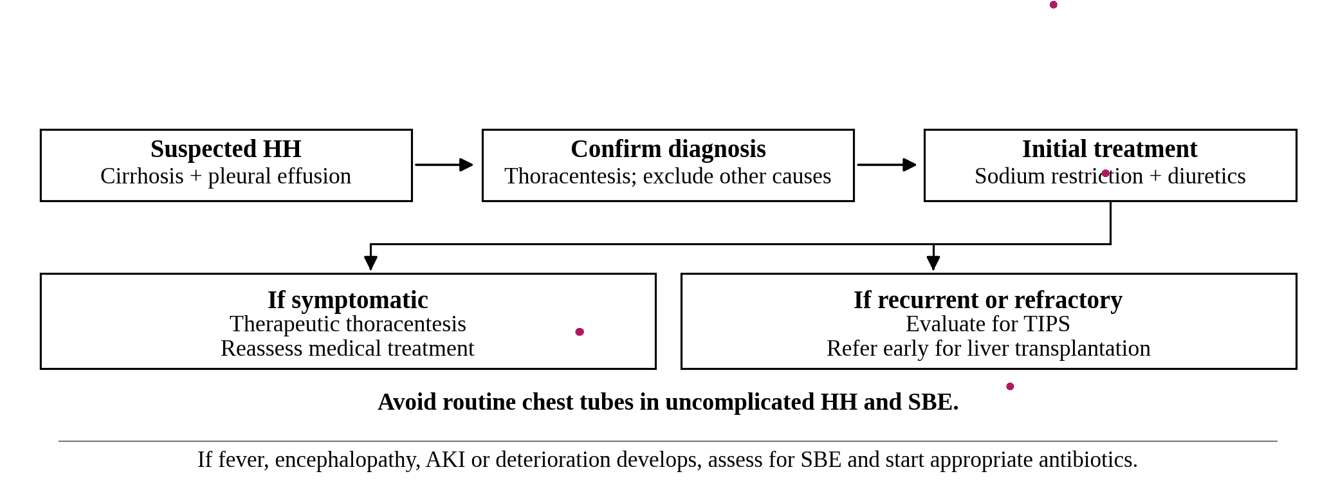
Figure 2. Stepwise diagnostic and therapeutic approach. SBE, spontaneous bacterial empyema; TIPS, transjugular intrahepatic portosystemic shunt.
Spontaneous bacterial empyema
Spontaneous bacterial empyema (SBE), or spontaneous bacterial pleuritis, is infection of HH in the absence of adjacent pneumonia. Suspect SBE in patients with fever, pleuritic pain, encephalopathy, acute kidney injury, sepsis, or otherwise unexplained deterioration; concurrent ascites or spontaneous bacterial peritonitis is not required. A pleural fluid polymorphonuclear leukocyte count >250 cells/mm³ in the appropriate setting warrants empiric antibiotics, selected according to acquisition setting and local resistance patterns. Chest imaging helps exclude pneumonia. Repeat pleural sampling after approximately 48 hours may be useful if the patient fails to improve. Routine chest tubes are not indicated for SBE, although frank pus or a secondary pleural infection requires separate drainage assessment.
When should liver transplantation be considered?
The diagnosis of HH should prompt consideration of transplantation, with early referral for recurrent or refractory disease. Its associated mortality can exceed the risk suggested by the calculated Model for End-Stage Liver Disease (MELD) score.
What does current MELD exception guidance require?
HH is not a standardized MELD exception. Under the July 1, 2025 OPTN/HRSA adult review guidance, a nonstandard exception may be considered in candidates with chronic, medically refractory, recurrent HH when TIPS has failed or is contraindicated and infectious and malignant causes have been excluded. The document guides National Liver Review Board decisions; it is not OPTN policy.
Table 1. Documentation for adult MELD exception review in refractory hepatic hydrothorax (July 2025 OPTN/HRSA guidance).
| Criterion | Documentation for review |
| Clinical setting | Chronic, recurrent, medically refractory HH; failed or contraindicated TIPS; infection and malignancy excluded. |
| Pleural drainage | At least 1 L removed on four separate occasions within 6 weeks. Record each date and volume; include provider or RN witness attestation if a catheter is in place. |
| Portal-hypertensive origin | Transudative or portal-hypertension-related fluid, supported by ascites or an albumin gradient ≥1.1 g/dL.* |
| Exclude alternative causes | Echocardiogram without heart failure; negative pleural culture or cell count and negative cytology, with dates. |
| Exception score | Candidates meeting the guidance should be considered for MMaT − 3, subject to board review. |
| Maintenance | Document four ≥1-L drainages in the preceding 6 weeks; update documentation every 90 days. |
*The 2025 guidance reverses the terms in its wording (“pleural albumin-serum albumin gradient”); clinically, SPAG is calculated as serum albumin minus pleural albumin. MMaT, median MELD at transplant.
Take-home points
- HH is a portal-hypertensive effusion, usually right-sided, and can occur without obvious ascites.
- For a new or unexplained effusion, diagnostic thoracentesis helps confirm the cause and detect infection.
- Treat initially with sodium restriction and diuretics; use thoracentesis for symptoms and consider TIPS and transplantation early in refractory cases.
- Avoid routine chest tubes in uncomplicated HH and SBE; use the current OPTN/HRSA criteria when requesting MELD exceptions.