Normal Liver Histology 101
Basic Architecture
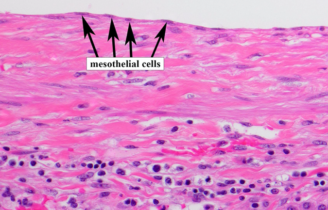
The Capsule
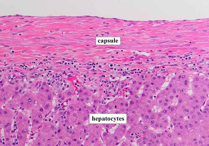
The outer surface of the liver is composed of a fibrous / connective tissue capsule (Figure 1). The outermost surface is covered by a thin layer of mesothelial cells that arises from the peritoneum (Figure 2).
The Parenchyma
The capsule surrounds the liver parenchyma, which is composed of hepatocytes. Hepatocytes are large polygonal cells with eosinophilic (pink) cytoplasm, round nuclei, and prominent nucleoli (Figure 1). Hepatocytes are discussed in more detail in later sections.
Organization: Lobules & Acini
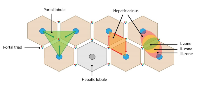
Microscopically, the organization of liver parenchyma can be represented by three different schematics: (1) the classic (hepatic) lobule, (2) the portal lobule, and (3) the hepatic acinus.
The Lobules
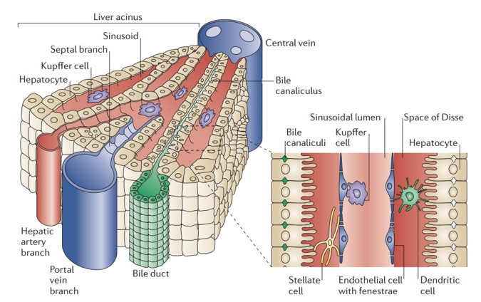
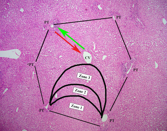
Classic (hepatic) lobules are based upon blood flow. They are organized as “hexagons,” with the central vein (CV) in the center of the lobule and portal tracts (PTs) at the periphery of the lobule (corners of the hexagon) (Figures 3 and 4). The PT consists of the hepatic artery (HA), portal vein (PV), and bile duct (BD). Blood enters the liver via the PV and HA, drains into the CV, and exits the liver via the hepatic vein to the inferior vena cava.
Portal lobules are based upon bile flow. They are organized as “triangles,” with a PT at the center of the lobule and CVs at the periphery of the lobule (corners of the triangle) (Figure 3). Hepatocytes produce bile that drains from the bile canaliculi > canals of Hering > bile ductules > interlobular bile ducts > intrahepatic ducts > right and left hepatic ducts > common hepatic duct > joins with cystic duct to form the common bile duct > duodenum). The flow of bile is in the opposite direction of blood flow (Figure 4).
The Hepatic Acinus (Acinus of Rappaport)
The hepatic acinus is based upon blood perfusion throughout the liver parenchyma. The acinus is organized as a “diamond,” with PTs at the corners of the short axis and CVs at the corners of the long axis (Figure 3). The acinus is arbitrarily divided into three zones based upon perfusion. Zone 1 receives the most oxygenated blood and nutrients, while zone 3 receives the least.
Key Structures
The Portal Tract
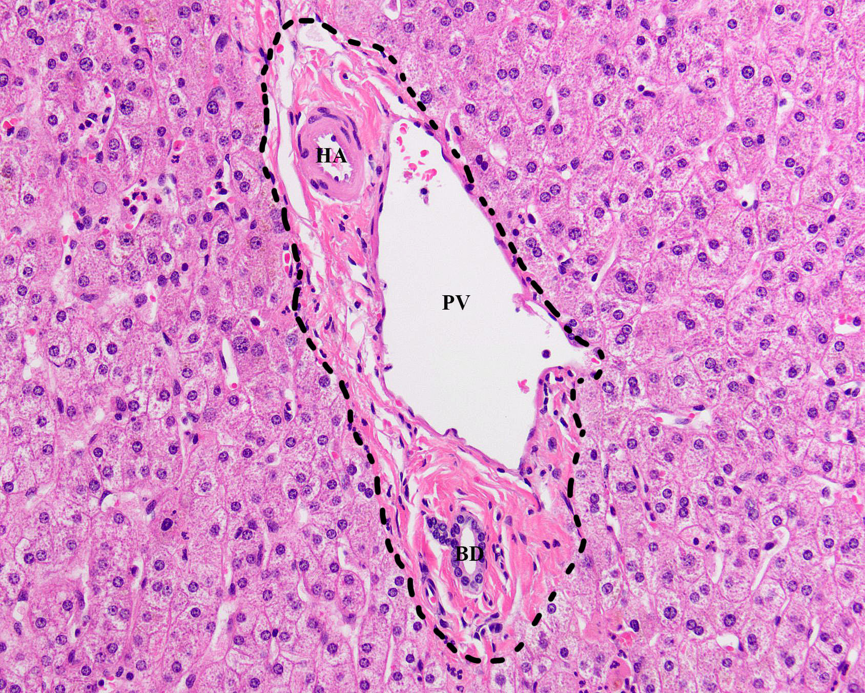
The PT consists of the HA, PV, and BD (Figure 5). The HA and PV are lined by endothelial cells. The HA is small with thick walls, while the PV is larger with thin walls. The BD is roughly the same size as the hepatic artery and is lined by a single layer of cuboidal (square-shaped) epithelium. Occasionally, lymphatic ducts may also be seen. The structures in the portal tract are surrounded by fibrous connective tissue. Although it is normal to see a few scattered inflammatory cells (ex. lymphocytes), no neutrophils or plasma cells should be present. The single layer of hepatocytes immediately adjacent to the portal tract delineates the limiting plate.
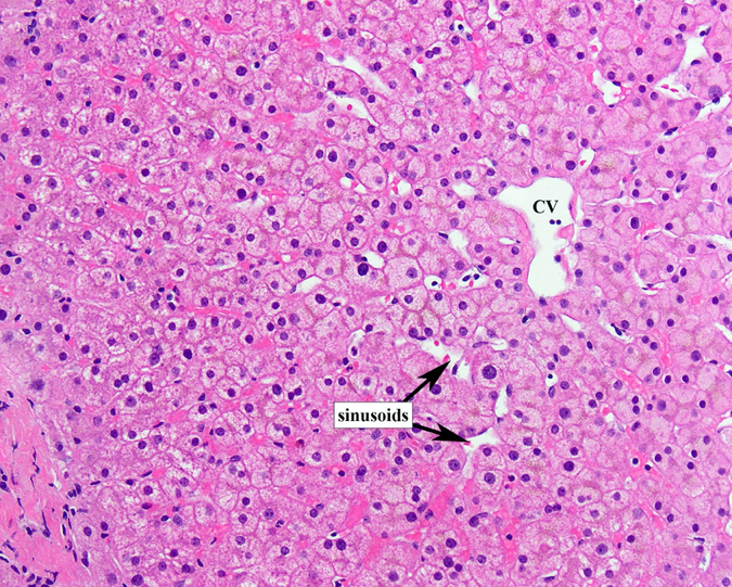
The Central Vein
The CV is lined by a single layer of endothelial cells and drains blood coming from the PTs via sinusoids. Plates of hepatocytes radiate outward from the CV toward the PTs (Figure 6).
Parenchyma
Hepatocytes
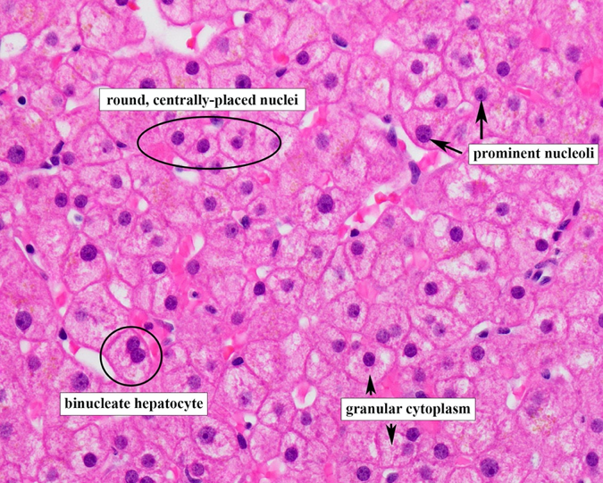
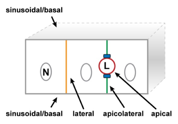
Hepatocytes are polygonal cells (20-30µm) with abundant granular eosinophilic (pink) cytoplasm, centrally placed round to ovoid nuclei, and prominent nucleoli (Figure 7). Normal hepatocytes can also show binucleation and pleomorphism (differences in size). Hepatocytes are arranged in plates that are 1 cell thick. Hepatocyte plasma membranes are separated into sinusoidal, canalicular, and apicolateral domains. The sinusoidal (basal) domain contains microvilli and faces the space of Disse, while the canalicular (apical) domain forms the bile canaliculus. The apicolateral domain is between the sinusoidal and canalicular domains (Figure 8).
Sinusoids
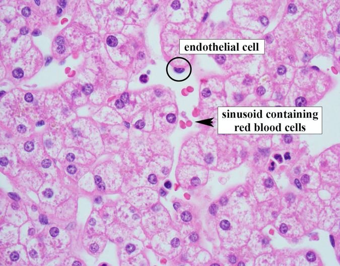
Hepatocytes are separated by sinusoids (Figures 9 and 10), which are spaces lined by fenestrated endothelial cells that transport blood from the PTs to the CVs. Sinusoids contain macrophages called Kupffer cells.
Space of Disse
The space of Disse (SoD) is the space between the endothelium and the hepatocytes (Figure 9), which transports lymphatic fluid to lymphatic capillaries in the portal tract and contains reticulin fibers. The SoD also contains stellate (Ito) cells that store lipids (vitamin A) and are involved in fibrinogenesis.