Why do patients with chronic liver disease get osteoporosis?

Why do patients with chronic liver disease get osteoporosis?
Case:
A 57-year-old woman with compensated cirrhosis from primary biliary cholangitis (PBC) presents to the emergency room after a ground level fall and is found to have a mildly displaced femoral neck fracture. She undergoes operative repair that is uncomplicated, but her hospital course is prolonged by immobility with development of a pressure ulcer and new decompensation with ascites and encephalopathy. She is ultimately discharged to a skilled nursing facility for rehabilitation.
Patients with chronic liver disease are twice as likely to suffer an osteoporotic fracture versus the general population. Hepatologists overlook the need to screen for low bone density in high-risk patients and consider fracture prevention as an important part of our care for all patients with chronic liver disease. This post will outline 1) why patients with liver disease develop low bone density and 2) the screening and management principles for osteoporosis that we should be comfortable with as hepatologists and 3) the natural history of bone mineral density in liver transplant recipients.
Introduction:
Hepatic osteodystrophy describes the spectrum of metabolic bone disease that can occur in patients with liver disease that encompasses osteopenia, osteoporosis, and osteomalacia. Osteoporosis is a loss of bone mass and weakening of the bone structure which predisposes patients to fractures. Risk factors for osteoporosis in the general population include advanced age, female gender, smoking, alcohol use, being underweight, early menopause (<45 years old), and treatment with steroids (Table 1).
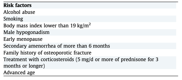
Table 1: Risk factors for development of osteoporosis in chronic liver disease. Figure adapted from the EASL guidelines for nutrition in chronic liver disease.
Patients with liver disease experience many of these baseline risk factors as well as other physiologic changes that make chronic liver disease an independent risk factor for bone density loss. It is estimated that 30% of patients with chronic liver disease also have osteoporosis. Patients with cholestatic liver disease, specifically PBC and primary sclerosing cholangitis (PSC) are the most effected, with the incidence of osteoporosis in PBC ranging from 20-44% in cross-sectional studies.
Fractures are prevalent for patients with chronic liver disease as are post-fracture complications such as infection, bleeding, and improper wound healing. Several studies have shown that patients with liver disease who experience a fracture have a higher mortality rate than matched controls (Figure 1).
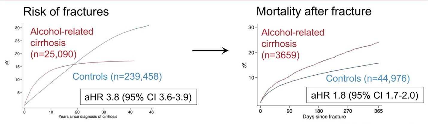
Figure 1: A retrospective cohort study using the Swedish National Patient Registry found patients with alcohol-related cirrhosis had a 4-fold higher risk of fracture as compared to matched controls. Patients with cirrhosis who experienced a fracture experienced 2-fold higher mortality rate.
Pathophysiology
Normal bone remodeling involves both bone breakdown or resorption by osteoclasts and bone formation by osteoblasts. Bone density loss results when the balance between these two processes is disrupted.
How does cholestasis lead to osteoporosis?
The link between bone density loss and cholestasis is incompletely understood, but several mechanisms have been explored. In the general population, osteoporosis is most common in post-menopausal women since estrogen inhibits osteoclasts, thus a decrease in estrogen levels leads to an increase in osteoclast activity and progressive bone resorption. Conversely, many of the factors driving osteoporosis in cholestasis and liver disease in general do so through anti-anabolic factors that decrease bone formation (Figure 2).
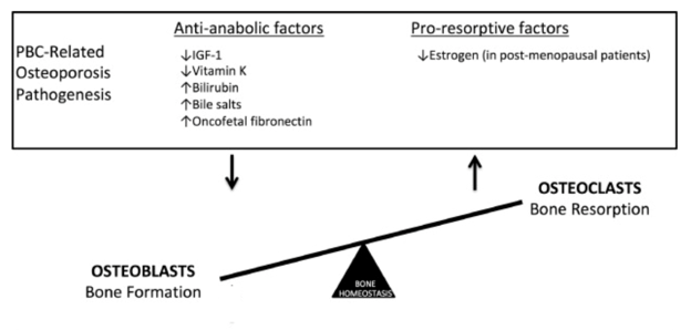
Figure 2: The proposed pathophysiology of bone density loss in primary biliary cholangitis (PBC). Osteoporosis in PBC develops due to factors that decrease normal bone formation. This is distinct from bone density loss in post-menopausal women which is driven by an increase in bone resorption due to a decrease in estrogen-mediated osteoclast apoptosis. Post-menopausal women with PBC are affected by both processes and are the most at risk for bone loss and fractures. Figure adapted from Danford et al.
Patients with cholestatic liver disease experience an abnormal transit of bile and bile acids from the liver into the small intestine, causing fat malabsorption and deficiencies in fat-soluble vitamins including vitamins D and K. Vitamin D3 is also synthesized in the skin, a process that is inhibited in patients with jaundice. This leads to lower levels of active vitamin D metabolites (eg: 1,25-hydroxyvitamin D3) and a downregulation of bone formation by osteoblasts as well as a decrease in uptake of calcium from the GI tract. Unabsorbed fat in the GI tract in patients with fat malabsorption binds dietary calcium and also leads to decreased calcium absorption.
Vitamin K is important in the synthesis of proteins that stimulate osteoblast activity and bone formation. A small randomized, controlled trial of patients with PBC saw a lower rate of bone mineral density loss in patients treated with daily vitamin K supplementation compared to controls. There is still insufficient evidence to recommend vitamin K supplementation for this indication, but this study supports the theory that vitamin K is integral to maintaining bone density.
A cholestatic state with elevated serum bilirubin and bile acids has also been shown to decrease bone formation through decreasing osteoblast viability and differentiation.
Why do patients with cirrhosis get osteoporosis?
Cytokines
Patients with cirrhosis experience an increase in cytokines such as interleukin (IL)-1, IL-6 and TNF-alpha that stimulate osteoclast activity and contribute to increased bone resorption. Patient with chronic liver disease also have lower levels of insulin-like growth factor (IGF)-1, an important stimulator of osteoblast activity that is synthesized by the liver. The lower levels of IGF-1 are likely due to hepatocyte dysfunction causing resistance to growth hormone signaling and decreased IGF-1 production.
Vitamin D
Vitamin D deficiency is common in patients with cirrhosis due to poor nutritional intake of vitamin D3. Synthesis of vitamin D3 in the skin requires exposure to sunlight, and patients with advanced liver disease, especially at northern latitudes, may not get adequate sunlight exposure to maintain healthy vitamin D levels. Vitamin D3 is converted in the liver to 25- hydroxy-vitamin D, a step that is limited in patients with advanced liver disease.
Hypogonadism
Hypogonadism is present in 90% of men with cirrhosis and can contribute to low bone density. Testosterone influences bone health directly through binding to androgen receptors on osteoblasts and promoting bone formation. Testosterone is converted to estrogen by aromatase, thus a decrease in testosterone also contributes to a loss of estrogen mediated inhibition of bone resorption by osteoclasts.
Alcohol
Heavy alcohol use has an indirect effect on bone density by causing an increase in inflammatory cytokines (eg IL-6 and TNF-alpha), vitamin D deficiency, and hypogonadism which disrupt normal bone turnover through the mechanisms described above. Ethanol is also directly toxic to osteoblast mediated bone formation with several studies showing a dose dependent decrease in osteoblast activity with exposure to alcohol. Alcohol causes an increase in reactive oxygen species and oxidative stress, which affects the cell signaling that governs differentiation of osteocyte progenitor cells into osteoblasts and osteoclasts. In patients with heavy, chronic alcohol use, these progenitor cells differentiate into adipocytes rather than osteoblasts, leading to an increase in bone marrow fat content rather than healthy bone formation.
Which patients with liver disease should be screened for low bone density?
Assessment of bone mineral density is done primarily with dual energy X-ray absorptiometry (DEXA) of the lumbar spine and proximal femur. AASLD and EASL guidelines agree that patients with cholestatic liver disease (PBC and PSC), autoimmune hepatitis with risk factors such as significant steroid exposure, and patients under evaluation for liver transplant should be screened for decreased bone density with a DEXA scan (Table 2). The AASLD has not extended the recommendation of a bone density assessment to all patients with cirrhosis, whereas EASL does recommend a DEXA scan for all patients with cirrhosis regardless of etiology.
It is important to note that the presence of ascites falsely lowers bone density readings on DEXA scans especially for the lumbar spine. This may contribute to an over diagnosis of osteoporosis in patients with cirrhosis and ascites. It is recommended that patients with ascites undergo a paracentesis prior to their DEXA scan to improve the accuracy of the results.
| Patient Group | Timing of first DEXA scan | Screening interval |
| Primary biliary cholangitis (PBC) | At diagnosis | Every 2 years |
| Primary Sclerosing Cholangitis (PSC) | At diagnosis | Every 2-3 years |
| Autoimmune hepatitis (AIH) | When risk factors are present (prolonged steroid use, postmenopausal status, history of low-trauma fracture, age >65 for women or >70 for men) | Every 2-3 years of continuous glucocorticoid treatment |
| Patients under evaluation for liver transplant | During evaluation | |
| Liver transplant recipients | When on a stable immunosuppression regimen, ~6-12mo post-transplant | For patients with osteoporosis/osteopenia, repeat every 1-2 years until bone density has stabilized |
Table 2: Summary of which patients with liver disease should be screened for low bone density with a dual-energy X-ray absorptiometry (DEXA) scan based on AASLD guidelines for the management of autoimmune hepatitis, primary biliary cholangitis, primary sclerosing cholangitis, liver transplant candidate evaluation, and non-graft related complications of liver transplant.
What are effective treatments for osteoporosis in patients with liver disease?
The cornerstone of osteoporosis treatment and prevention is optimizing nutrition and engaging in regular resistance exercise training. The recommendations around nutrition and exercise for patients with chronic liver disease are detailed in other LFN posts on nutrition and frailty, thus we will focus on interventions specifically for osteoporosis.
Vitamin D and Calcium
Vitamin D and calcium supplementation is recommended for all patients with PBC and for patients with chronic liver disease who have osteoporosis or osteopenia. Recommended calcium intake is 1000-1200mg per day along with 400-800 IU of vitamin D3, with high dose vitamin D repletion for patients with serum levels of 25-hydroxyvitamin D less than 30 mg/ml. Of note, the effectiveness of vitamin D and calcium supplementation in improving bone mineral density and fracture risk chronic liver disease has never been proven.
Bisphosphonates:
Bisphosphonates such as alendronate, cyclic etidronate, risendronate, and zolendronic acid prevent bone resorption and are effective in treating osteoporosis in post-menopausal women. Several small studies have shown efficacy of bisphosphonates in slowing bone density loss in patients with PBC; however, pooled analysis from a systematic review and meta-analysis of osteoporosis treatments in PBC showed no statistically significant improvement in bone mineral density or decrease in fractures with bisphosphonate treatment. The lack of efficacy for standard osteoporosis therapies in the PBC population may be since most treatments, including bisphosphonates, are decrease bone resorption, while patients with liver disease experience mostly a decrease in bone formation as the driver of their bone density loss.
There are no randomized, controlled trials of osteoporosis treatment in patients with cirrhosis. A target trial emulation study of Medicare enrollees with cirrhosis and osteoporosis did find a lower rate of fractures in those treated with a bisphosphonate. There is theoretical concern that bisphosphonates that might increase the risk of variceal bleeding in patients with portal hypertension given their association with pill esophagitis; however, recent retrospective studies have supported their safety in this population.
Denosumab
Denosumab is a monoclonal antibody that binds to a receptor on osteoclasts suppressing their activity and decreasing bone resorption. It is a subcutaneous injection administered once every 6 months. Nonrandomized studies of denosumab in patients with chronic liver disease show a positive effect on bone mineral density without significant safety concerns or adverse events. Studies in older adults have shown denosumab treatment is associated with improvement in sarcopenia as well as bone density, highlighting the connection between sarcopenia and osteoporosis and the potential of interventions in treating both.
Beyond the therapies discussed above, there are many other treatments for osteoporosis now available, in particular anabolic agents that support bone growth (ie: teriparatide, abaloparatide, and romosozumab). Further studies of these therapies in the cirrhosis population are needed to show safety and efficacy particularly in prevention of osteoporotic fractures.
What happens to bone density before and after liver transplant?
Osteoporosis is prevalent in liver transplant recipients and associated with a high rate of fracture, estimated at 25 to 35% within the first year after transplant. Bone density decreases in the first 3 to 6 months post-transplant likely due to immobility and steroid exposure. With improvement in nutritional status and mobility as well as decreased immunosuppression, bone mineral density stabilizes and may increase starting at 6 to 12 months post-transplant.
The AST/AASLD guidelines on the care of patients after orthotopic liver transplant (OLT) recommends that liver transplant recipients undergo a DEXA scan once they are on a stable immunosuppression regimen, which is typically around 6-months post-transplant. Calcium and vitamin D supplementation are recommended for all patients post-transplant with the goal of maintaining 25-hydroxyvitamin D levels above 30ng/mL. Recommendations for management of osteoporosis and osteopenia post-transplant largely mirror the recommendations for the general population as studies specifically in the post-OLT population have been small and heterogeneous. A systematic review and meta-analysis of osteoporosis treatment post-OLT did find that treatment with bisphosphonates significantly decreased fracture risk without significant adverse events. There is insufficient evidence to support the use of bisphosphonates prophylactically to prevent bone loss and fractures in this population.
As hepatologists, we have the opportunity to partner our primary care and endocrinology colleagues to ensure that post-OLT patients are evaluated for low bone density within 2 years after transplant and that they receive the standard of care for osteoporosis treatment. As post-OLT bone density recovery can take months to years even with pharmacologic treatment, much of the approach for fracture prevention in the immediate post-OLT period should focus on physical therapy for fall prevention.
Key Points:
- Compared with the general population, patients with chronic liver disease, particularly those with primary biliary cholangitis (PBC), have an increased risk of osteoporosis, osteoporotic fractures, and fracture-related morbidity and mortality.
- Factors contributing to bone density loss in cholestatic liver disease include deficiencies in fat soluble vitamins like vitamin D and K and the inhibitory effect of cholestasis on osteoblast function and bone formation.
- Patients with cirrhosis experience bone density loss due to factors such as an increase in inflammatory cytokines, vitamin D deficiency, hypogonadism, sarcopenia, and alcohol use.
- Patients with PBC, PSC, autoimmune hepatitis with risk factors for bone density loss, and all patients under evaluation for liver transplant should be screened for osteoporosis with a DEXA scan.
- The presence of ascites falsely lowers bone density readings on DEXA scan, thus it is recommended to perform a DEXA scan soon after a paracentesis for patients with significant ascites.
- Bisphosphonates are safe and can be effective in treating osteoporosis and preventing fractures in patients with chronic liver disease as well as post-liver transplant.
- Bone density decreases in the first 3-6 months after liver transplant and patients in the early post-transplant period are at a particularly high risk of fracture.