Dyspnea After Liver Transplant: What’s Taking His Breath Away?

Case

A 66-year-old male with a past medical history of primary biliary cholangitis s/p orthotopic liver transplant 10 months ago, ESRD s/p kidney transplant 2 months ago, presents with shortness of breath and dyspnea on exertion. His symptoms have gradually worsened over 2 weeks, and he is now short of breath walking across a room. He denies fevers, chills, and sick contacts. He is compliant with his medications and currently on prednisone, mycophenolate mofetil, and tacrolimus for immunosuppression. Liver chemistries are within normal limits and chest imaging shows a large right pleural effusion and right anterior chest wall mass. What is the most likely underlying diagnosis?

What is the most likely underlying diagnosis?

Correct Answer:

Post-transplant lymphoproliferative disorder

The most likely diagnosis for this patient is post-transplant lymphoproliferative disorder (PTLD). The increased immunosuppression the patient received after his recent kidney transplant led to the development and progression of PTLD, manifesting as a chest wall mass and pleural effusion that caused his shortness of breath. This diagnosis was ultimately confirmed with pleural fluid studies and a tissue biopsy. Keep reading to learn about risk factors for PTLD, and how it can present with a variety of symptoms.

Symptoms and Diagnosis of PTLD

The clinical presentation of PTLD is highly variable. Due to the systemic nature of the disease, a high index of suspicion must be maintained when considering PTLD in a patient’s differential diagnosis. Patients may present with constitutional symptoms, such as weight loss, fevers or night sweats, and swollen lymph glands, or more specific complaints, including abdominal pain, gastrointestinal bleeding, or sore throat. Physical exam signs include the following: lymphadenopathy, hepatosplenomegaly, oral ulcers, and focal neurologic deficits. Histopathologic examination is ultimately necessary to confirm the diagnosis of PTLD. Remember, the incidence of PTLD after 5 years post liver transplant is 1.1%.  This schematic outlines the important elements of PTLD diagnosis:

Figure 1 - Approach to Diagnosis of PTLD after Liver Transplant

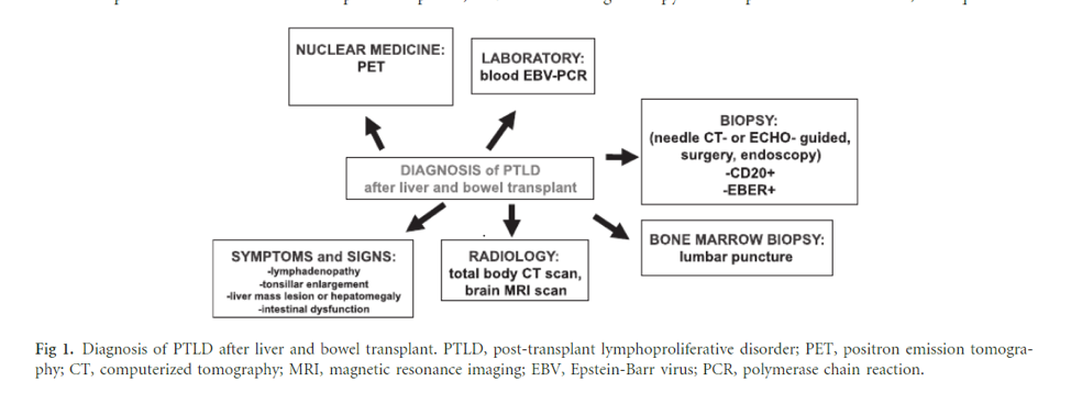
Image taken from: Managing the Challenge of PTLD in Liver and Bowel Transplant Patients. BJH 2015; 169:157-172.

Back to the case

The patient underwent a thoracentesis with two liters of pleural fluid removed. Fluid analysis revealed an exudative effusion by Light’s criteria and showed 23% unclassified cells. Give the location  of the mass, a triple phase liver MRI was obtained. This showed retrocaval, mesenteric, and retroperitoneal lymphadenopathy, along with a large mass indenting upon the liver. Fungal (blastomycosis, histoplasmosis, coccidiomycosis) and viral studies (CMV, EBV) were negative. A CT-guided biopsy of the right chest wall mass was then performed, with histopathology showing a high grade B-cell lymphoma compatible with B-cell lymphoma.

Given the findings of a chest wall mass and LAD, a hepatic hydrothorax (HH) is unlikely to account for the patient’s symptoms. Additionally, in most cases of HH, the pleural fluid is transudative by Light’s criteria (Need a refresher on HH? Review our recent LFN post here). Lung cancer could account for his presentation, however the extent of his lymphadenopathy and the pathology results confirm a lymphoma, compatible with PTLD. Similarly, the patient is at risk for a disseminated infection such as histoplasmosis or tuberculosis, but the biopsy is not consistent with infection. He also lacked signs and symptoms typical of infections, such as fevers, chills, and night sweats. 

Risk Factors for PTLD

The biggest risk factors for PTLD are immunosuppression, EBV infection, and age. Both increased intensity and duration of immunosuppression increase the risk for formation of PTLD. Calcineurin inihibitors, anti-thymocyte globulin, and azathioprine are the agents with the greatest risk for causing PTLD.

PTLD is more than twenty times more common in EBV seronegative patients than EBV seropositive patients. Immunosuppression leads to T cell downregulation, which can promote EBV infection, lymphocyte proliferation, and ultimately PTLD.

Management of PTLD

The first step in treatment for PTLD is reduction in immunosuppression. Expert recommendations include stopping anti-proliferative agents such as azathioprine and mycophenolate and reducing calcineurin inhibitor therapy by 30-50%. Studies show a 45% response rate, 37% complete response rate, and 17% relapse rate with this strategy. 

The gold standard treatment regimen is R-CHOP: rituximab, cyclophosphamide, doxorubicin, vincristine, and prednisone. Below are the summarized results of the PTLD-1 trial (Figure 2), which notably demonstrates a high response rate to R-CHOP with a median overall survival of 6.6 years, as well as an algorithm for PTLD treatment (Figure 3).

Figure 2 - R-CHOP treatment regimens in patients with PTLD from the PTLD-1 trial

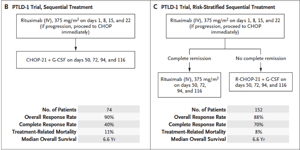
Taken from:  Post-transplant Lymphoproliferative Disorder in Adults. N Engl J Med 2018; 378:549-562. CHOP: cyclophosphamide, doxorubicin, vincristine, prednisone; PTLD: post-transplant lymphoproliferative disorder

Figure 3 - Approach to Management of PTLD

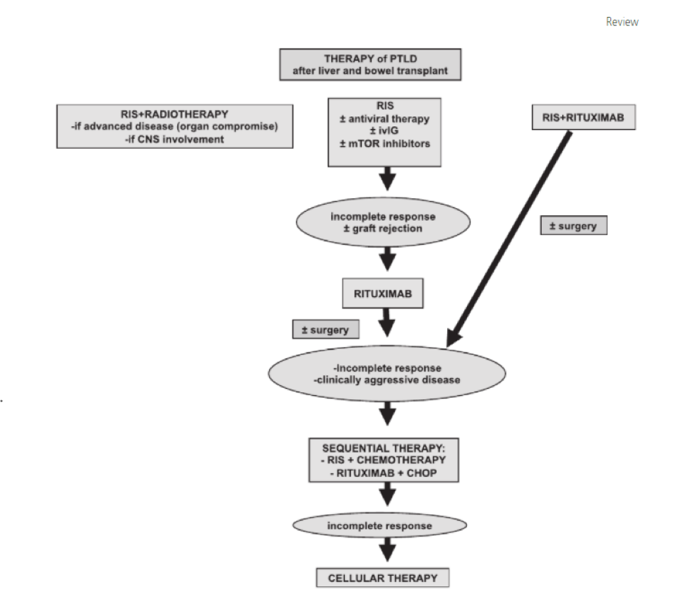
Taken from: Managing the Challenge of PTLD in Liver and Bowel Transplant Patients. BJH 2015; 169:157-172.

Back to the case

After the biopsy was obtained, the patient’s immunosuppression was reduced—his tacrolimus was halved and cellcept was discontinued. The patient was discharged with follow up in oncology clinic, where the chemotherapy regimen R-CHOP was promptly started, with a robust clinical response.